
In recent years, the integration of artificial intelligence and minimally invasive concepts has propelled the advancement and application of robotic technology in neurosurgery. As a key player in this field, the medical robot has revolutionized stereotactic procedures by enhancing precision, reducing trauma, and streamlining surgical workflows. This study focuses on evaluating the clinical accuracy of a domestically developed neurosurgical medical robot in frameless stereotactic surgeries. The medical robot, specifically designed for neurosurgical applications, combines computer-aided planning, visual tracking, and robotic manipulation to achieve high-precision targeting. Through retrospective analysis of clinical cases, we aim to quantify accuracy errors and identify factors influencing these errors, thereby optimizing the performance of the medical robot in diverse surgical scenarios.
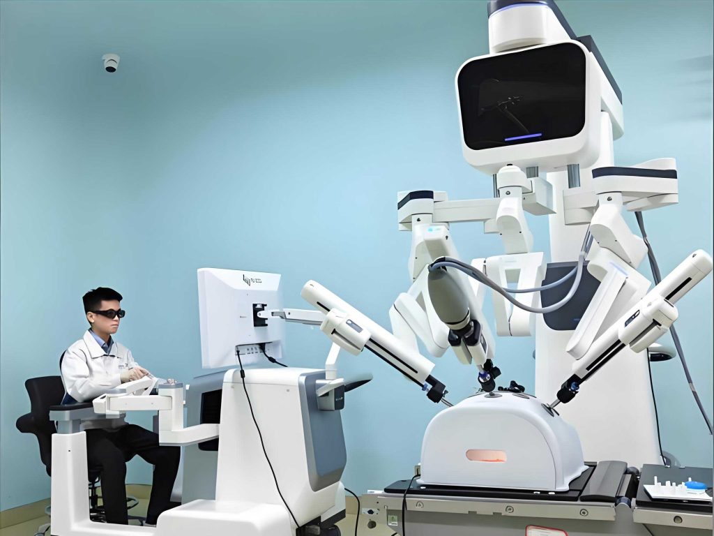
The medical robot system employed in this study features a six-degree-of-freedom robotic arm capable of precise three-dimensional movements, enabling the deployment of various instruments such as biopsy needles, electrodes, and drainage tubes. Its frameless design eliminates the need for traditional stereotactic frames, reducing patient discomfort and setup time. The core components include a planning platform for imaging fusion and trajectory simulation, a visual guidance system for real-time tracking, and an automated robotic arm for instrument positioning. This integration allows for seamless translation of preoperative plans into surgical actions, making the medical robot a versatile tool for procedures like biopsy, electrode implantation, and hematoma drainage.
To assess the accuracy of the medical robot, we conducted a retrospective review of 26 cases undergoing frameless stereotactic surgery with robotic assistance. These cases encompassed a range of procedures: 10 biopsies, 9 electrode implantations for stereo-electroencephalography (SEEG) and lesion coagulation, and 7 hematoma drainage surgeries. Based on the rigidity of the puncture instruments, target points were categorized into three groups: biopsy-coagulation group (14 targets), SEEG electrode implantation group (13 targets), and drainage tube insertion group (9 targets). This grouping allows for comparative analysis of accuracy errors attributable to instrument material properties, a critical aspect in evaluating the medical robot’s performance.
The surgical protocol with the medical robot involved several standardized steps. Preoperatively, patients underwent CT or MRI scans for target planning. The medical robot’s software was used to fuse imaging data and simulate trajectories. After patient registration using fiducial markers, the robotic arm autonomously positioned itself to the planned entry point. Following skin incision and burr hole creation, instruments were inserted along the guided path. Postoperative CT scans were obtained and fused with preoperative plans to measure deviations. Accuracy error was defined as the three-dimensional Euclidean distance between the planned target point and the actual target point, computed using the medical robot’s integrated software. This metric provides a comprehensive assessment of the medical robot’s targeting precision.
Statistical analysis was performed to compare accuracy errors across groups. Data were normally distributed and presented as mean ± standard deviation. Due to unequal variances, the Welch test was used for overall comparison, followed by Dunnett’s T3 test for pairwise comparisons. A significance level of P < 0.05 was adopted. Additionally, we derived formulas to model error propagation and conducted sensitivity analyses to explore the impact of various factors on the medical robot’s accuracy.
The results revealed distinct accuracy profiles for each group, highlighting the influence of instrument rigidity. The biopsy-coagulation group, utilizing rigid instruments like biopsy needles and coagulation electrodes, exhibited the smallest error: 1.330 ± 0.566 mm. In contrast, the SEEG electrode implantation group showed a larger error of 2.404 ± 0.878 mm, while the drainage tube insertion group had the highest error at 6.188 ± 4.103 mm. Statistical comparisons confirmed that the biopsy-coagulation group’s error was significantly lower than both the SEEG and drainage groups (P < 0.05), whereas no significant difference existed between the SEEG and drainage groups (P > 0.05). These findings underscore the medical robot’s capability when paired with rigid instruments, but also expose challenges with flexible tools.
| Group | Number of Targets | Accuracy Error (mm, mean ± SD) | Instrument Material |
|---|---|---|---|
| Biopsy-Coagulation | 14 | 1.330 ± 0.566 | Rigid (biopsy needle, coagulation electrode) |
| SEEG Electrode Implantation | 13 | 2.404 ± 0.878 | Flexible (depth electrode) |
| Drainage Tube Insertion | 9 | 6.188 ± 4.103 | Soft (silicon drainage tube) |
To further investigate error sources, we measured the entry point accuracy for the drainage group, which yielded 1.412 ± 0.415 mm, comparable to the biopsy-coagulation group’s target error (P = 0.713). This suggests that the large target error in drainage cases primarily stems from post-insertion displacement of soft tubes rather than inherent inaccuracies of the medical robot’s targeting system. The medical robot’s core positioning mechanism thus demonstrates high precision, but instrument behavior post-insertion introduces variability.
Error analysis in medical robot-assisted surgery involves multiple components. We can model the total error (E_total) as a combination of independent factors:
$$ E_{\text{total}} = \sqrt{E_{\text{registration}}^2 + E_{\text{mechanical}}^2 + E_{\text{instrument}}^2 + E_{\text{patient movement}}^2} $$
where E_registration is the error from patient-to-image registration, E_mechanical is the robotic arm’s kinematic error, E_instrument is error due to instrument flexibility or deflection, and E_patient movement is error from intraoperative head displacement. For the medical robot, registration errors arise from fiducial marker identification and potential marker shift, typically ranging from 0.5 to 1 mm. Mechanical errors, influenced by the robotic arm’s calibration and backlash, are minimized through high-precision encoders and repeatability tests. Instrument-related errors, however, dominate in flexible tool scenarios, as seen in our data.
The deflection of flexible instruments can be described by beam bending theory. For a cylindrical instrument inserted into brain tissue, the lateral displacement (δ) at the target depth (L) due to tissue resistance can be approximated by:
$$ \delta = \frac{F L^3}{3EI} $$
where F is the lateral force exerted by tissue, E is the Young’s modulus of the instrument material, and I is the area moment of inertia. For rigid instruments (high E), δ is negligible, whereas for soft materials (low E), δ becomes significant, explaining the increased errors in electrode and drainage tube groups. This mathematical insight emphasizes the need for instrument stiffness optimization in medical robot applications.
| Comparison | Mean Difference (mm) | 95% Confidence Interval | P-value |
|---|---|---|---|
| Biopsy-Coagulation vs. SEEG | -1.074 | [-1.892, -0.256] | 0.004 |
| Biopsy-Coagulation vs. Drainage | -4.858 | [-8.123, -1.593] | 0.021 |
| SEEG vs. Drainage | -3.784 | [-7.891, 0.323] | 0.068 |
Beyond instrument material, other factors contribute to accuracy errors in medical robot surgeries. Patient movement after registration, though mitigated by head immobilization with molded pillows, remains a concern; even slight shifts can degrade accuracy. Adapter tolerances between the robotic arm and instruments introduce coaxial errors, particularly angular deviations that amplify over distance. The medical robot’s software algorithms for image fusion and trajectory planning also have inherent limitations, though continuous updates have improved fidelity. Comparative studies with other medical robot systems, such as ROSA and Neuromate, show similar challenges, with reported electrode placement errors averaging 2-4 mm, underscoring the ubiquity of these issues in robotic neurosurgery.
To enhance the medical robot’s performance, several strategies are proposed. For flexible instrument delivery, the use of rigid guiding cannulas could reduce deflection, as evidenced by the entry point accuracy in drainage cases. Improved registration techniques, such as surface matching or intraoperative imaging, may lower E_registration. Additionally, real-time compensation algorithms based on force feedback could dynamically adjust trajectories to account for tissue deformation. The medical robot’s integration with intraoperative MRI or CT could further validate and correct targeting during surgery, pushing accuracy toward sub-millimeter levels.
The clinical implications of these findings are substantial. High accuracy in biopsy and coagulation procedures enables precise lesion targeting, improving diagnostic yield and therapeutic outcomes. For epilepsy surgery, electrode placement errors of 2-3 mm may still suffice for SEEG recordings, but finer precision could enhance focus localization. In hematoma drainage, while absolute accuracy is less critical due to catheter repositioning capability, reducing initial placement error minimizes tissue damage and accelerates recovery. Thus, the medical robot’s role extends beyond mere navigation; it facilitates tailored approaches based on procedural demands.
Future directions for medical robot development involve artificial intelligence augmentation. Machine learning models could predict instrument deflection based on preoperative imaging and tissue properties, allowing for preemptive path correction. Autonomous robotic systems might adapt trajectories in real-time using optical tracking or impedance sensing. Moreover, expanding the medical robot’s repertoire to include endoscopic or laser-assisted procedures could broaden its impact in minimally invasive neurosurgery. Collaborative efforts between engineers and clinicians will drive these innovations, ensuring that the medical robot evolves to meet complex surgical needs.
In conclusion, our analysis demonstrates that the neurosurgical medical robot achieves high positioning accuracy, particularly with rigid instruments, where errors average around 1.3 mm. Flexible instruments, however, introduce significant post-insertion errors due to material deflection, highlighting a key limitation. By addressing error sources through technical refinements and instrument design, the medical robot can further solidify its role as a precision tool in frameless stereotaxy. The continuous evolution of medical robot technology promises to enhance surgical outcomes, reduce invasiveness, and expand the frontiers of neurosurgical care, making it an indispensable asset in modern operating rooms.