
In modern neurosurgery, the pursuit of precision and minimal invasiveness has driven significant technological advancements. Among these, the integration of medical robot systems into surgical practice represents a paradigm shift, enabling more accurate, stable, and less traumatic procedures. As a neurosurgeon involved in this evolving field, I have witnessed firsthand the transformative impact of medical robots, particularly in stereotactic biopsy procedures for intracranial lesions. Stereotactic biopsy is a critical technique for diagnosing brain tumors and other pathological conditions, but traditional frame-based methods have limitations such as patient discomfort, restricted operative freedom, and challenges in pediatric cases. The advent of frameless stereotactic systems coupled with medical robots addresses these issues, offering enhanced flexibility and accuracy. This article delves into the application and precision analysis of a specific medical robot system in stereotactic biopsy, focusing on factors influencing accuracy, such as cerebrospinal fluid (CSF) loss during surgery. Through a detailed study, we explore how medical robots can optimize surgical outcomes and pave the way for future innovations in neurosurgery.
The medical robot system under discussion is a frameless stereotactic platform that integrates surgical planning, registration, and robotic arm manipulation. This medical robot utilizes a six-degree-of-freedom intelligent mechanical arm to guide instruments, such as biopsy needles, with high precision. Preoperatively, imaging data like CT and MRI are imported into the planning software, allowing for 3D reconstruction of intracranial structures and lesion delineation. The system facilitates trajectory planning while avoiding critical vasculature and functional areas. During surgery, the medical robot employs visual tracking to register fiducial markers dynamically, ensuring accurate spatial alignment. The robotic arm then positions the instrument to the planned target, minimizing human error. This medical robot has been deployed in various neurosurgical procedures, including tumor biopsy, hematoma drainage, and deep brain electrode implantation, demonstrating its versatility and reliability. In this study, we assess the target accuracy of this medical robot in stereotactic biopsy and investigate the effect of intraoperative CSF loss on precision, proposing a simple yet effective mitigation strategy.
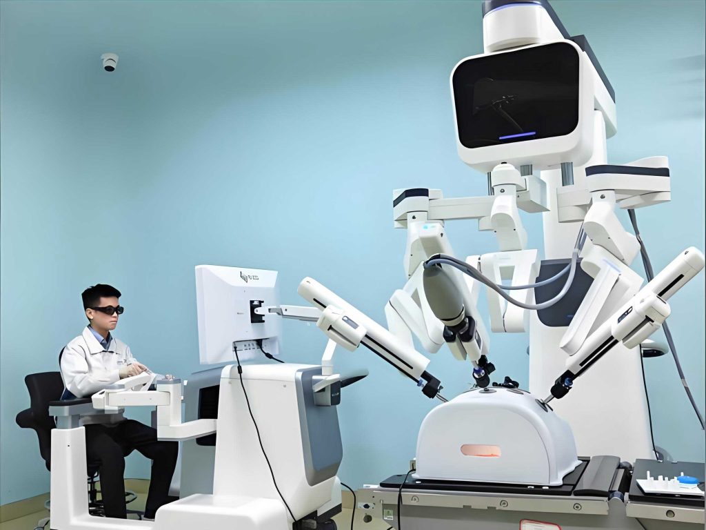
To evaluate the medical robot’s performance, we conducted a prospective analysis involving patients undergoing frameless stereotactic brain biopsy. All procedures were performed at our institution, adhering to ethical guidelines and obtaining informed consent. Patients were selected based on clinical indications, such as indeterminate infiltrative lesions on imaging, deep-seated or multifocal pathologies, or contraindications to open craniotomy. A total of 64 patients were enrolled, with ages ranging from 21 to 78 years and a mix of frontal, temporal, thalamic, and multifocal lesions. Each patient underwent a single biopsy from one target site. Preoperatively, five skin-affixed fiducial markers were applied, followed by thin-slice CT scanning from the vertex to the nasal tip. MRI data were fused with CT images in the medical robot’s planning platform to define the target coordinates and trajectory. The surgical workflow involved general anesthesia, head fixation, automated registration via the medical robot’s camera system, and verification of accuracy. After skin incision and burr hole creation, the dura was opened, and the arachnoid and pia mater were coagulated and incised. At this point, patients were divided into two groups based on preference: the experimental group (36 patients) received immediate application of a medical biological glue (porcine fibrin sealant) to seal the burr hole and prevent CSF loss, while the control group (28 patients) had no intervention. The medical robot’s arm then guided the biopsy needle to the target for tissue sampling. Postoperatively, a CT scan was obtained, and the images were fused with preoperative data to measure the Euclidean distance between the planned target and the actual biopsy site, representing the target accuracy error. Statistical analysis was performed using independent samples t-test to compare errors between groups, with significance set at p < 0.05.
The demographic and clinical characteristics of the patients are summarized in Table 1. Both groups were comparable in terms of age, gender distribution, and lesion locations, ensuring minimal bias in the accuracy assessment. The medical robot facilitated all procedures without major complications, such as intracranial infection or significant hemorrhage. Minor intralesional bleeding occurred in five cases, likely due to vascular injury during biopsy, and resolved with conservative management. The overall target accuracy error for the medical robot-assisted stereotactic biopsy was calculated as the mean Euclidean distance in three-dimensional space. Let the planned target coordinates be \( (x_p, y_p, z_p) \) and the actual target coordinates be \( (x_a, y_a, z_a) \). The error \( E \) is given by:
$$ E = \sqrt{(x_a – x_p)^2 + (y_a – y_p)^2 + (z_a – z_p)^2} $$
This formula was applied to each case, and the results are presented in Table 2. The overall mean error was 1.510 mm with a standard deviation of 0.636 mm, indicating high precision of the medical robot system. However, when comparing the experimental and control groups, a significant difference emerged. The experimental group, with burr hole sealing using medical biological glue, had a mean error of 1.207 mm (SD = 0.470 mm), while the control group had a mean error of 1.899 mm (SD = 0.614 mm). The t-test yielded a statistic of -5.108 with a p-value of 0.001, confirming that the reduction in error in the experimental group was statistically significant. This suggests that minimizing CSF loss during surgery enhances the accuracy of the medical robot-assisted procedure.
| Variable | Experimental Group (n=36) | Control Group (n=28) |
|---|---|---|
| Age (years), mean ± SD | 44.3 ± 11.4 | 47.8 ± 10.8 |
| Gender, male/female | 22/14 | 18/10 |
| Lesion Location: | ||
| Frontal lobe | 7 | 3 |
| Temporal lobe | 10 | 5 |
| Thalamus | 7 | 9 |
| Multifocal | 12 | 11 |
| Group | Mean Error (mm) ± SD | 95% Confidence Interval |
|---|---|---|
| Overall (n=64) | 1.510 ± 0.636 | [1.350, 1.670] |
| Experimental (n=36) | 1.207 ± 0.470 | [1.047, 1.367] |
| Control (n=28) | 1.899 ± 0.614 | [1.656, 2.142] |
The precision of the medical robot system can be further analyzed by considering sources of error. In frameless stereotaxy, errors may arise from registration inaccuracies, patient movement, instrument flexibility, and anatomical shifts. The medical robot’s design mitigates some factors through rigid head fixation, tight instrument适配, and dynamic tracking. However, brain shift due to CSF loss remains a challenge, as evidenced by our results. The medical biological glue used in the experimental group acts as a sealant, forming a barrier that reduces CSF egress and stabilizes intracranial contents. The glue, composed of fibrinogen and thrombin, polymerizes rapidly upon contact with tissue fluids, creating a semi-transparent gel that hardens within minutes. Its properties allow easy penetration by the biopsy needle without interference, making it ideal for this application. The reduction in error from 1.899 mm to 1.207 mm highlights the importance of controlling CSF loss, which can otherwise lead to brain displacement and target misalignment. This finding aligns with previous studies on brain shift in stereotactic procedures, underscoring the need for integrated strategies in medical robot-assisted surgery.
Beyond CSF management, the medical robot’s accuracy is influenced by procedural details. For instance, the choice of surgical position and burr hole location can affect CSF dynamics. Positioning the entry point at a higher cranial site may minimize fluid loss, and the medical robot’s planning software allows optimization of trajectory based on patient posture. Additionally, the rigidity of the biopsy needle compared to softer instruments like drainage catheters contributes to lower inherent error in biopsy procedures. The medical robot system achieves sub-millimeter precision in ideal conditions, but real-world factors necessitate compensatory measures. The error formula above can be extended to account for systematic biases, such as registration offset. If we denote the registration error as a vector \( \vec{R} = (r_x, r_y, r_z) \), the total error \( E_t \) might be modeled as:
$$ E_t = \sqrt{(x_a – x_p + r_x)^2 + (y_a – y_p + r_y)^2 + (z_a – z_p + r_z)^2} $$
However, in our study, the medical robot’s verification step ensured registration accuracy, so the primary variable was CSF-related shift. The medical robot’s ability to fuse pre- and post-operative images for error calculation itself demonstrates its utility in quality control and continuous improvement.
The discussion of medical robot applications in neurosurgery extends beyond biopsy to broader implications. Medical robots like this system enhance surgical planning through 3D visualization and simulation. For example, the trajectory planning algorithm can incorporate risk scores based on proximity to vessels or eloquent areas, optimizing safety. The medical robot’s mechanical arm provides steadier instrument holding than human hands, reducing tremor and fatigue. This is particularly beneficial in deep-seated lesions where minimal deviation is critical. Moreover, the medical robot supports tele-surgery and training, potentially expanding access to expert care. In our experience, the medical robot has reduced procedure time and improved patient outcomes, with no major complications attributed to the system. The integration of medical robots into routine practice requires attention to factors like cost, training, and workflow adaptation, but the precision gains justify investment. Future developments may include real-time MRI guidance, artificial intelligence for trajectory optimization, and enhanced haptic feedback, further solidifying the role of medical robots in neurosurgery.
In conclusion, medical robots represent a significant advancement in stereotactic neurosurgery, offering high precision and minimal invasiveness. Our study focused on a medical robot-assisted frameless biopsy system, showing an overall target accuracy error of 1.510 mm, which was significantly improved to 1.207 mm by sealing the burr hole with medical biological glue to prevent CSF loss. This underscores the importance of mitigating brain shift during medical robot procedures. The medical robot’s capabilities in planning, registration, and execution make it a valuable tool for diagnosing intracranial pathologies, with applications expanding to other interventions. As medical robot technology evolves, we anticipate further reductions in error margins and broader adoption across neurosurgical centers. The medical robot is not just a tool but a partner in achieving surgical excellence, embodying the principles of precision medicine. Continued research into factors affecting accuracy, such as CSF dynamics and instrument design, will enhance the performance of medical robots, ultimately benefiting patients through safer and more effective treatments.