
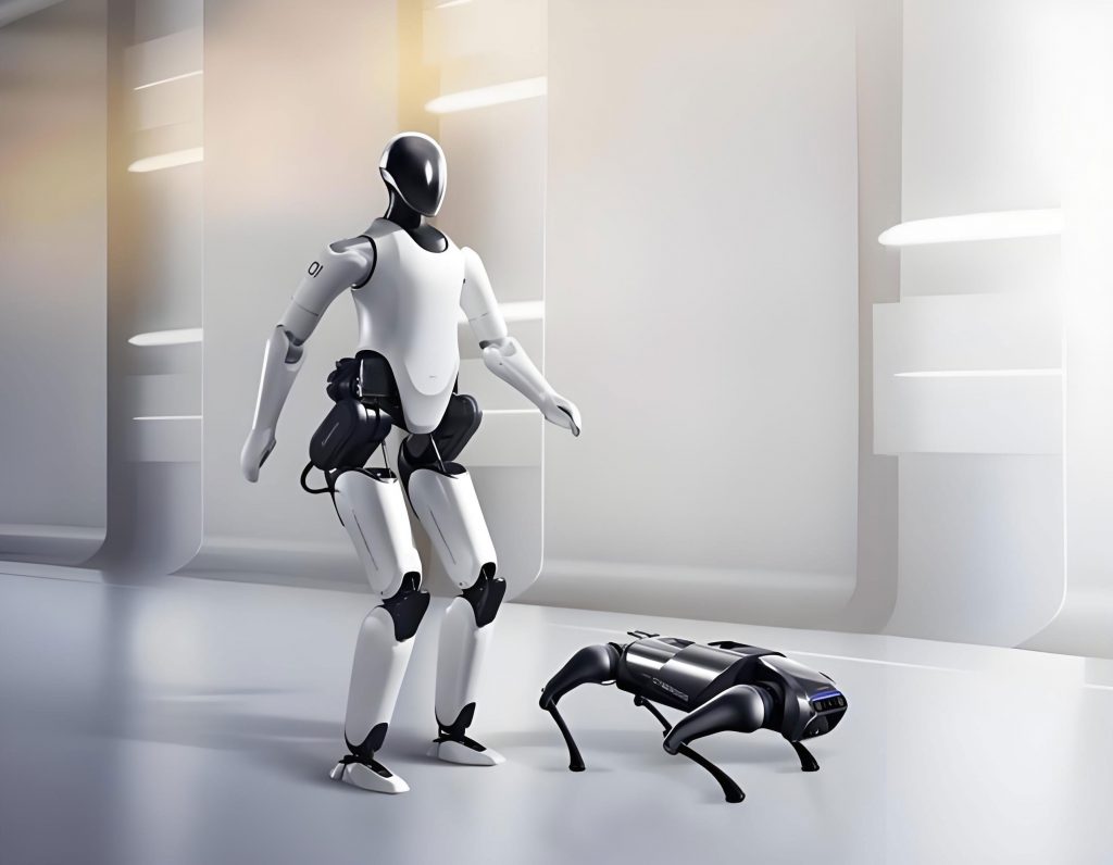
As a researcher deeply immersed in the field of medical robotics, I find the evolution of bionic robots for internal diagnostics to be one of the most fascinating and impactful technological journeys. A bionic robot is fundamentally an apparatus constructed from mechanical components and electronic chips, programmed to emulate the locomotive characteristics of living organisms. From snake and canine mimics to avian and crustacean forms, these bionic robots have proven their utility in military reconnaissance, eldercare assistance, and intelligence gathering. Within this broad spectrum, a particularly compelling niche is the miniaturization of bionic robots to simulate small organisms like insects, worms, and bees. This line of inquiry is gaining significant, specialized attention. The application of micro-robots in clinical diagnosis and treatment promises not only enhanced reliability and authenticity of examinations but also a drastic reduction in patient discomfort due to the minuscule size and high intelligence of Micro-Electro-Mechanical Systems (MEMS). This is especially crucial for operations within the constrained, non-structured environments of the human body. For instance, capsule endoscopy, with its minimal invasiveness, absence of toxic side effects, and potential for active control and imaging, represents the inevitable future of robotic endoscopic technology.
The passive wireless capsule endoscope that emerged in recent years marked a revolutionary step toward truly non-invasive internal examination. Roughly the size of a standard fish oil capsule, it transmits images wirelessly, can be swallowed, passes through the digestive tract via natural peristalsis, and is eventually excreted. Its small size and passive nature cause no trauma to the intestinal lining and result in zero patient discomfort. However, this very passivity is its Achilles’ heel. The capsule cannot be moved forward or backward at will, preventing physicians from re-examining areas of interest. Its transit is too rapid in the esophagus, its view is limited in the stomach, and it cannot inspect behind folds in the colon. Consequently, its primary effectiveness is currently confined to the small intestine. Furthermore, dependence on button-cell batteries limits operational life to approximately 7-8 hours, rendering it ineffective for patients with slower gastrointestinal transit times.
The limitations of passive capsules necessitated the development of active, controllable systems. The first logical step was magnetic manipulation. By embedding a small magnet within the capsule and using an external magnetic field for remote control, magnetically guided capsules were created. These have found success in painless gastric examinations, leveraging the stomach’s larger cavity. However, the intricate, lengthy, and fold-rich environment of the intestines poses a formidable challenge for effective magnetic control. To address this, researchers turned to a more classic bionic robot inspiration: legged locomotion. The development of a legged capsule robot, featuring slender, flexible, insect-like legs, enables stable walking and driving within the intestinal environment. This approach is currently undergoing clinical trial investigations, representing a significant leap toward fully navigable intestinal bionic robots.
Beyond legged motion, another profound source of inspiration comes from organisms like earthworms and tapeworms, which excel in soft, slippery environments using peristaltic or inchworm-like locomotion. Mimicking these gaits is key to achieving physiologically compatible and safe propulsion within the special non-structured environment of human lumens. The goal is to find a traction method that causes minimal disruption, aligning perfectly with medical objectives. Researchers have extensively explored the inchworm bionic structure, initially successful in industrial pipeline robots. While the environments share similarities, direct transplantation to clinical use is impossible. The core principle of inchworm motion involves having at least one segment firmly anchored (or “berthed”) to the surface while other segments elongate or contract longitudinally. Effective berthing is paramount.
In rigid industrial pipes, berthing is achieved by pressing mechanisms against the wall to generate sufficient friction. In contrast, intestinal tissue is a soft, highly deformable, viscoelastic biological surface with a very low coefficient of friction. A robot that deforms too little will slip; one that deforms too much risks exceeding physiological tolerance and causing injury. This dichotomy has led to the investigation of three primary support mechanisms for inchworm-style bionic robots:
| Support Mechanism | Principle | Advantages | Disadvantages & Risks |
|---|---|---|---|
| Pneumatic (Airbag) | Inflation of a flexible bladder to contact the lumen wall. | Good flexibility, conforms to tissue. | Large deformation may damage tissue; control complexity. |
| Mechanical (Clamping) | Alternating extension/retraction of rigid or semi-rigid gripping elements. | Positive anchoring, good control. | Risk of localized trauma or perforation; less compliant. |
| Adhesive (Vacuum) | Creating negative pressure to suction the lumen wall to the robot’s surface. | Potentially gentle, distributed force. | Low adhesion force due to mucosal layer and villi; sealing challenge. |
The effectiveness of these mechanisms hinges on the fundamental physics of friction. The maximum anchoring force $F_{anchor}$ before slippage occurs is governed by:
$$ F_{anchor} = \mu \cdot N $$
where $\mu$ is the coefficient of static friction between the robot’s contact material and the mucosal tissue, and $N$ is the normal force exerted by the support mechanism (e.g., clamping force, inflation pressure, or suction pressure). For a bionic robot to move effectively, the driving force from its elongation/contraction must be less than $F_{anchor}$ for the stationary segment. The low $\mu$ of wet mucosal tissue is therefore a central design challenge, often requiring a trade-off between increasing $N$ and the risk of tissue damage.
Parallel to the berthing challenge is the quest for suitable micro-actuators. The confined space demands extreme miniaturization, with each actuator type presenting a unique set of trade-offs in terms of force, displacement, speed, and power efficiency.
| Actuator Type | Working Principle | Advantages for Micro Bionic Robots | Disadvantages & Challenges |
|---|---|---|---|
| Shape Memory Alloy (SMA) | Material deformation upon heating (phase change). | High force-to-weight ratio, relatively large stroke, simple structure, easily miniaturized. | Low energy efficiency, slow cooling/cycle time, requires heat dissipation management. |
| Pneumatic | Controlled air pressure inflating micro-chambers or bellows. | High force, inherent softness/flexibility, good safety profile. Power source can be external. | Requires a tether (supply tube), control latency, potential for leakage. |
| Piezoelectric | Material deformation under applied electric field. | High precision, fast response, high stiffness, silent operation. | Extremely small strain/displacement, requires high voltage, often needs amplification mechanisms. |
| Micro-Motor | Electromagnetic rotation converted to linear motion. | Mature technology, high efficiency, easy control, continuous rotation. | Requires complex miniaturized gearboxes for force conversion, potential for electromagnetic interference. |
The kinematics of an inchworm-style bionic robot can be modeled for a simple two-segment, three-node configuration. Let $L_1$ and $L_2$ be the lengths of the front and rear segments (contractile/dilative elements), and $x_f$, $x_m$, $x_r$ be the positions of the front, middle, and rear nodes. One step cycle involves:
1. Rear Anchoring & Front Extension: Rear segment anchors ($x_r$ fixed). Front segment extends from $L_1$ to $L_1 + \Delta L$. The middle node advances: $x_m’ = x_m + \Delta L$.
2. Front Anchoring & Rear Contraction: Front segment anchors ($x_m’$ fixed). Rear segment contracts, pulling the rear node forward: $x_r’ = x_r + \Delta L$.
The net displacement of the robot’s body per cycle, $\delta$, is ideally equal to the actuator stroke $\Delta L$, but in practice is reduced by slippage $\epsilon$:
$$ \delta = \Delta L – \epsilon $$
where $\epsilon$ is directly related to the insufficiency of the anchoring force $F_{anchor}$ during each phase.
In contrast to the inchworm gait, the earthworm (peristaltic) gait offers a different paradigm for a bionic robot. Instead of distinct anchored and moving segments, a peristaltic robot generates a traveling wave of contraction and expansion along its body. A multi-segment model is essential here. For a robot with $n$ segments, the state of the $i$-th segment can be defined by its strain $\varepsilon_i(t)$. A coordinated control wave $C(z, t)$ propagates from tail to head, where $z$ is the body coordinate:
$$ C(z, t) = A \cdot \sin(2\pi (ft – z/\lambda)) $$
where $A$ is the amplitude of contraction, $f$ is the wave frequency, and $\lambda$ is the wavelength. The strain in a segment at position $z_i$ is then:
$$ \varepsilon_i(t) \propto C(z_i, t) $$
This wave interacts with the frictional environment. A common model assumes anisotropic friction: a higher static friction coefficient $\mu_s$ when the segment is radially expanded (simulating setae in earthworms), and a lower kinetic friction coefficient $\mu_k$ when it is contracted and sliding. The net propulsive force $F_{prop}$ over one wavelength can be approximated by integrating the differential friction forces:
$$ F_{prop} \approx n_{anchor} \cdot (\mu_s – \mu_k) \cdot \langle N \rangle $$
where $n_{anchor}$ is the number of expanded segments anchored at a given time, and $\langle N \rangle$ is the average normal force from radial expansion. This model highlights the critical role of friction coefficient modulation rather than just force magnitude. While effective on level surfaces, the peristaltic bionic robot‘s ability to climb slopes or resist retrograde peristalsis becomes highly sensitive to the precise ratio $\mu_s / \mu_k$. Achieving this anisotropy reliably on wet, soft tissue remains a significant hurdle.
The design of any lumen-traversing bionic robot is a complex optimization problem within severe spatial constraints. The primary objective is to maximize propulsive efficiency and diagnostic capability within a cylinder of diameter $D_{max}$ (often < 15mm) and length $L_{max}$. This involves trade-offs described by a series of interrelated parameters:
| Design Parameter | Symbol | Constraint/Goal | Trade-off Relationship |
|---|---|---|---|
| Total Robot Diameter | $D$ | $D \leq D_{max}$ (Lumen diameter) | Limits actuator cross-section, battery size, electronics. |
| Actuator Stroke | $\Delta L$ | Maximize for speed. | Larger $\Delta L$ requires longer actuator or complex mechanics, increasing $L$. |
| Actuator Force | $F_a$ | $F_a > F_{drag} + F_{slope}$ | Higher $F_a$ typically requires more power or larger actuator volume. |
| Power Consumption | $P$ | Minimize for battery life. | $P \propto F_a \cdot v / \eta$, where $v$ is speed and $\eta$ is efficiency. |
| On-board Energy | $E$ | Maximize for procedure duration. | $E \propto \rho_{energy} \cdot D^2 \cdot L$, limited by biocompatible battery tech. |
| Diagnostic Payload Volume | $V_{payload}$ | Maximize for cameras, sensors, tools. | $V_{payload} \approx \frac{\pi}{4}D^2 L – V_{actuators} – V_{electronics} – V_{battery}$ |
The future of gastrointestinal bionic robots lies in overcoming these intertwined challenges. Material science will contribute through novel biocompatible, soft polymers with tunable surface properties to manage friction. Advanced micro-fabrication will enable more powerful and efficient micro-actuators. Energy harvesting from the biochemical environment or wireless power transfer could solve the endurance problem. Finally, the integration of artificial intelligence for semi-autonomous navigation and real-time diagnostic analysis (e.g., automated polyp detection) will transform the device from a simple camera capsule into an intelligent diagnostic bionic robot.
In conclusion, the journey from passive capsule to actively navigated bionic robot for gastrointestinal diagnostics encapsulates a remarkable convergence of biology, robotics, and medicine. While significant hurdles in actuation, anchoring, power, and control persist, they are of a nature that is foreseeable and surmountable with sustained interdisciplinary research. The ongoing development of legged, inchworm, and peristaltic bionic robots paves the way for a new era of truly non-invasive and comprehensive endoscopic examination. The widespread clinical adoption of these microscopic marvels will undoubtedly revolutionize early detection and treatment of gastrointestinal diseases, fundamentally altering the landscape of preventive healthcare and oncology outcomes.